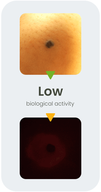
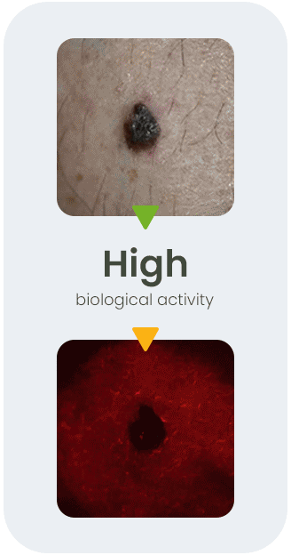


Fluorescent biotag is topically applied then wiped away

Handheld imager captures white light and fluorescent images

The software provides a probability score for tissue remodeling

Fluorescent Biotag
Binds to Receptors

Imager CapturesFluorescence

Software Reports the Presence of a Malignancy Biomarker
